
Mark Reed, DPM 714-528-FOOT
1275 Rose Drive Placentia, CA Suite 136
Melanie Reed, DPM 714-528-7777
1275 Rose Drive Placentia, Ca Suite 124
Podiatrists @ Placentia-Linda Foot & Ankle Group
BUNION DEFORMITY
CAUSES AND TREATMENT
Bunions and foot pain have been discussed in the medical podiatry literature for more than 100 years. The term "bunion" is derived from the Latin word for turnip. Bunions usually occur on both feet with one foot usually worse than the other. In western countries, bunions occur more in women due to the type of shoes women wear. In countries where men and women do not wear shoes, the incidence of bunions in men and women has been found to be the same. However, the person's inherited or developed tendency to walk flat- footed has been shown to be the primary cause of bunion deformities. In rare circumstances, neurological disorders, rheumatoid arthritis, and developmental deformities can also cause a bunion deformity.
Evaluation of a bunion deformity begins with obtaining a complete history and physical as well as obtaining X-rays of both feet. Typically, as a bunion deformity progresses, calluses form under the ball of the foot, a reddened area appears over the prominent first metatarsal head, lesser toe deformities appear, shoes no longer fit properly, degenerative arthritic joint changes occur and the cosmetic appearance of the foot deteriorates.
Treatment of a bunion deformity primarily involves two issues, correcting the underlying cause of the bunion deformity, and when required, surgically realigning the bunion deformity. In cases where the bunion deformity is mild to moderate and the patient is not in pain, custom molded foot orthotics are prescribed to help correct the weight bearing forces which usually cause the bunion deformity.
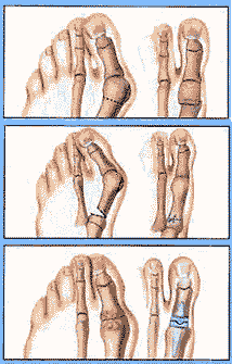 Surgical
realignment of a bunion deformity consists of a realignment of
the great toe and first metatarsal. In some cases where
arthritic joint degeneration has occurred, besides realigning
the joint deformity, the joint must also be fused or artificially
replaced. A bunion deformity actually involves two unique
deformities. The first is the widening of the knuckle joint of
the great toe from the knuckle joint of the second toe and is
called Metatarsus Premus Adductus. Metatarsus Premus Adductus
is measured by measuring the angle created by drawing a bisection
of the first and second metatarsal bones on a weight-bearing
AP X-ray. The angle of the first metatarsal to the second
metatarsal is called the 1-2 Inter-metatarsal Angle and usually
is normal if under 10 degrees. The second deformity is
the rotation of the great toe towards the second toe that is
called the Hallux Abductovalgus Deformity.
Surgical
realignment of a bunion deformity consists of a realignment of
the great toe and first metatarsal. In some cases where
arthritic joint degeneration has occurred, besides realigning
the joint deformity, the joint must also be fused or artificially
replaced. A bunion deformity actually involves two unique
deformities. The first is the widening of the knuckle joint of
the great toe from the knuckle joint of the second toe and is
called Metatarsus Premus Adductus. Metatarsus Premus Adductus
is measured by measuring the angle created by drawing a bisection
of the first and second metatarsal bones on a weight-bearing
AP X-ray. The angle of the first metatarsal to the second
metatarsal is called the 1-2 Inter-metatarsal Angle and usually
is normal if under 10 degrees. The second deformity is
the rotation of the great toe towards the second toe that is
called the Hallux Abductovalgus Deformity.
In evaluating a patient for surgical correction of their bunion deformity, ultimately, the different causes of the patient's Metatarsus Premus Adductus and Abductovalgus Deformity must be determined by the surgeon. Usually, the cause of the bunion deformity is solely due to widening of the first metatarsal away from other metatarsals. However, there are many anatomical reasons that a bunion deformity can be in existence and this is why a foot specialist should evaluate a bunion deformity prior to surgery. In correcting a bunion deformity, some doctors still cut off a portion of the knuckle joint of the great toe to narrow the appearance of the foot but do not realign the knuckle joint of the great toe. This type of narrowing procedure typically severely narrows the knuckle joint of the great toe and has been associated with numerous complications such as early degenerative joint arthritis. Typically, these narrowing procedures are temporary in giving any relief to the patient and other surgical procedures must be performed subsequently to realign the knuckle joint of the great toe. A second opinion should be obtained from a foot specialist if a doctor recommends a narrowing procedure that intends to narrow the knuckle joint of the great toe without realignment of the joint.
In discussing recovery expectations, in the hands of a good foot surgeon, for the patient with a mild to moderate bunion deformity that involves only the widening of the first metatarsal compared to the second metatarsal, the patient will have usually a two to four week recovery period before returning to shoes and can walk on the foot during recovery. For the patient with a severe bunion deformity that involves only the widening of the first metatarsal from the second metatarsal, surgery usually involves an eight to ten week period in which no weight bearing can occur.
In making a pre-operative evaluation of the patient, weight bearing X-rays must be evaluated to determine he appropriate procedures that need to be performed. The amount of angle between the first and second metatarsal is one of the main factors in determining which type of procedure is indicated and how much recovery time will be experienced by the patient. The angle between the first and second metatarsals is important because there is a point that the first metatarsal head can not be sided back gains the second metatarsal. When the first metatarsal is too wide to slide it over against the second metatarsal, a wedge procedure must be performed to swing the metatarsal over far enough to move the first metatarsal head against the second metatarsal head. In evaluating X-rays, there are actually 11 other X-ray measurements that are typically evaluated in deciding what is the correct procedure for correcting a bunion deformity.
Early medical intervention using custom molded functional foot orthotics can in many cases prevent bunion surgery or slow the progression of the deformity. The use of foot orthotics after bunion surgery is important in preventing the return of the bunion deformity. Please review the foot orthotic article for information on foot orthotics. There are other interventions that help such as using a silicon spacer to help align the joint to reduce stress to the ligaments of the joint.
The best approach in deciding what to do about a developing buinon is to get an X-ray early in the progression of the deformity to gain an understanding of what is the best intervention to either prevent the deformity from increasing in severity or correcting the deformity before permanent arthritic changes occur to the joint that can not be reversed with corrective surgery.